A precise and highly specialized method used to surgically remove skin cancer
MOHS Micrographic Surgery
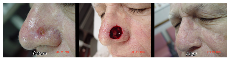
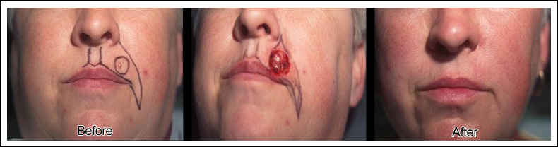
MOHS Micrographic Surgery is a precise and highly specialized method used to surgically remove skin cancer. The cancerous tissue is excised under local anesthesia and then immediately examined under microscopic vision. This proven method of skin cancer removal offers the greatest possible chance of a complete cure while preserving as much normal skin as possible.
MOHS Surgery: Why it’s Unique
There are several common methods currently used to examine cancerous tissue when removed from a patient and sent to a pathologist for margin evaluation. Bread loaf sectioning, cross sectioning, and peripheral sectioning involve the evaluation of vertical sectioned samples from the tissue submitted to a lab for processing. While these methods are commonly used, they only allow a physician to examine a very small percentage of the margins of most concern. In contrast, the MOHS technique allows for the skin cancer tissue to be processed in such a way that horizontal sections are obtained, which allows for the microscopic evaluation of the entire base of the removed tissue as well as the entire outside peripheral margin. Therefore, the most vital information required to determine whether all the cancer has truly been eliminated is obtained by this exacting technique—MOHS Micrographic Surgery.
MOHS Surgery - The Advantages
The goals in treating skin cancer are as follows:
- to offer a patient the highest chance of cure;
- to conserve as much healthy tissue as possible
- to minimize dysfunction or distortion
- to achieve the best cosmetic result
MOHS Micrographic Surgery can achieve these goals unlike any other available method, which is why it has become the gold standard for the removal of many of the most common types of skin cancers.
MOHS Micrographic Surgery is usually most beneficial and cost effective when used to treat skin cancers that have developed on delicate locations of one’s face and for skin cancers that have not responded to the more common methods of removal. The most common skin cancers that are treated in this manner are basal cell and squamous cell carcinomas.
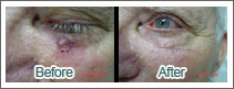
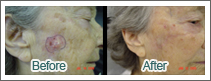
When skin cancers are discovered in locations close to one’s eyes, ears, lips, or nose, the MOHS technique allows a physician to conserve as much normal skin as possible, while also ensuring that the cancer is completely removed. More than any other technique used today, MOHS Micrographic Surgery helps minimize unnecessary distortion or dysfunction, which is of utmost importance in achieving the best cosmetic result.
In summary the MOHS removal method is superior to other methods in that the skin cancer that is removed is evaluated under microscopic vision immediately on site. The patient need not agonize while waiting days for the lab results, and in many instances, the wound resulting from the removal can be fixed the same day since the MOHS surgeon is confident that the cancer has been completely removed and thoroughly evaluated.
“Mohs micrographic surgery is considered the most effective technique
for treating many basal cell carcinomas (BCCs) and squamous cell
carcinomas (SCCs), the two most common types of skin cancer.”
- Skin Cancer Foundation
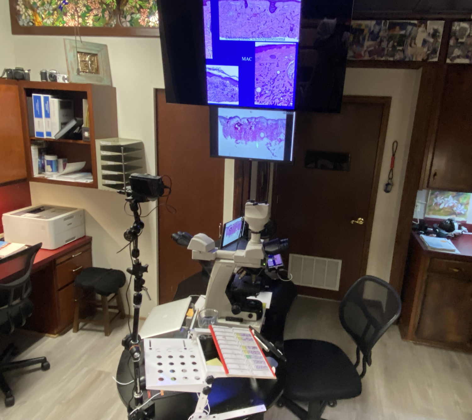
Skin Cancer Centre Laboratory
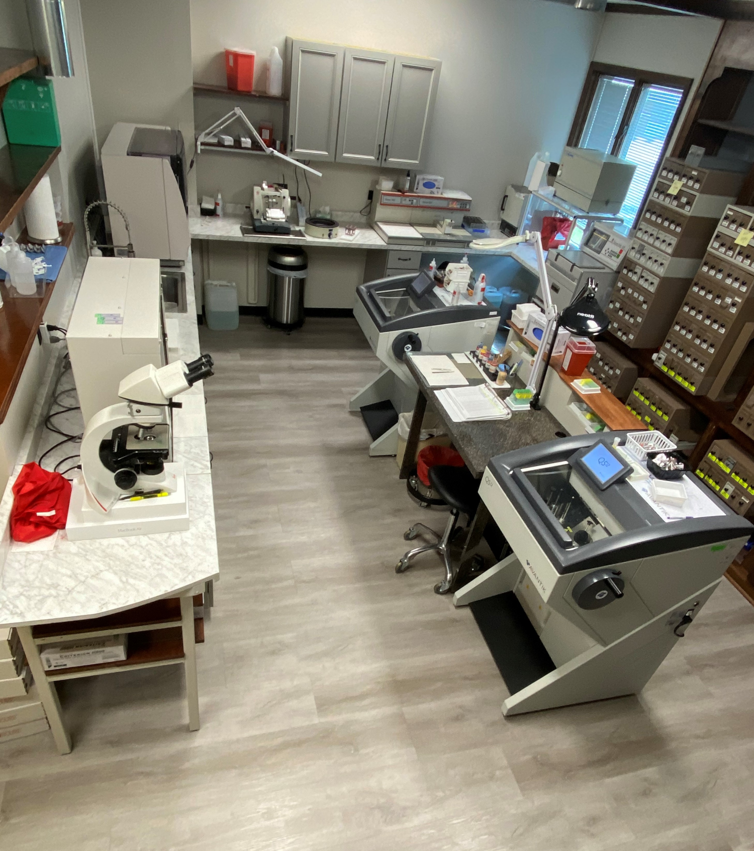
The Skin Cancer Centre's laboratory is an essential part of our practice. It allows us to process tissue in-house to deliver real-time diagnoses for our Mohs cases.
Thanks to our experienced and extremely efficient histotechnician, we are able to create slides from your tissue sample enabling microscopic evaluation almost immediately. This ensures that we conserve as much of your healthy tissue as possible while also giving our team the greatest chance of removing all of the cancer.